

How AI is Revolutionising Breast Cancer Screening Using Medical Imaging
Breast cancer is the most diagnosed cancer in women. According to the World Health Organization report in 2022, 2.3 million women were diagnosed with breast cancer, and it was also estimated that 7.3 million women are battling breast cancer globally. By 2040, the cases of breast cancer are expected to rise to 3 million new cases and 1 million deaths annually due to population growth and aging alone. Early detection is our best weapon against this disease; it can dramatically improve survival rates and reduce the need for extensive surgeries. But here’s the challenge: radiologists (doctors who specialise in interpreting medical images to diagnose and treat diseases) are overwhelmed, medical images are becoming increasingly complex, and in many parts of the world, there simply are not enough specialists to screen everyone who needs it.
That is where artificial intelligence (AI) comes in. It is a game-changer that’s transforming how we detect and diagnose breast cancer.
In our recently published research, we explored how deep learning (an advanced type of AI) can support doctors in detecting breast cancer more accurately and efficiently. Think of AI as a smart assistant that learns from thousands of medical images to help radiologists spot suspicious areas that might be easy to miss.
What We Discovered
Our research team recently completed the most comprehensive review to date of how AI is being used in breast cancer screening. We analysed 124 studies published between 2019 and 2024, focusing on three main types of medical imaging: mammograms (X-ray images of the breast), ultrasounds, and MRI scans.
The results are remarkable. AI systems are now matching and sometimes exceeding the performance of experienced radiologists in detecting breast cancer. Some systems are already being used in hospitals to flag potential cancers, reduce reading time, and even help less experienced doctors make better decisions.
How AI is Making a Difference
Catching Cancer Earlier: AI systems can identify suspicious areas in breast images with incredible accuracy. In some studies, these systems detected cancer cases that radiologists had initially missed, potentially saving lives through earlier intervention.
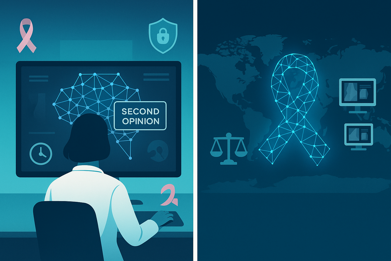
Supporting Doctors, Not Replacing Them: Contrary to fears about AI taking over, we found that the technology works best when it assists radiologists rather than replacing them. When doctors use AI as a “second opinion,” their diagnostic accuracy improves significantly, especially for less experienced radiologists.
Speeding Up Diagnoses: AI can analyse images in seconds rather than minutes, potentially reducing waiting times for patients and allowing doctors to see more patients each day.
Reducing False Alarms: Nobody wants to receive a cancer scare that turns out to be nothing. AI is helping reduce false positive results by up to 37% in some studies, meaning fewer unnecessary biopsies and less anxiety for patients.
Real-World Impact
Commercial AI systems are already being tested in hospitals and clinics around the world. These are not just laboratory experiments, they are real tools helping real doctors make better decisions about real patients. Some systems can automatically flag mammograms that are most likely to be normal, allowing radiologists to focus their time on cases that need the most attention.
What Radiologists Think
We also explored what doctors themselves think about AI in breast cancer screening. The good news? Most radiologists are optimistic about AI’s potential to improve patient care. However, they have important concerns:
· They want AI systems that can explain their decisions, not just give a “yes” or “no” answer
· They are concerned about data privacy and security
· They want to ensure AI systems work fairly for all patients, regardless of their background
· They emphasise that AI should enhance their capabilities, not replace their judgment
The Road Ahead
While the progress is exciting, we are not quite ready for fully automated breast cancer screening. Current AI systems still have limitations – they often work well in laboratory settings but need more testing in real-world clinical environments with diverse patient populations.
The future looks bright, though. We are moving toward AI systems that can:
· Work seamlessly with existing hospital technology
· Provide clear explanations for their recommendations
· Handle different types of medical images from various manufacturers
· Maintain patient privacy while continuously learning and improving
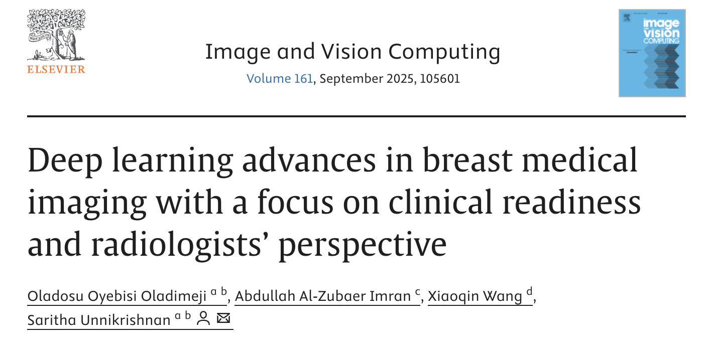
One challenge remains: most AI tools today are tested in single hospitals using limited data. For AI to work well for all women across different ages, ethnicities, and breast types, it needs to be trained and tested on diverse, large-scale datasets. Fairness, safety, and privacy must also be top priorities.
The good news is that we are moving in the right direction. With continued collaboration between AI developers, clinicians, and patients, intelligent tools could soon become a standard part of breast cancer screening, making the process faster, more accurate, and accessible to more people.
What This Means for Patients
For women facing breast cancer screening, this research brings hope. In the coming years, you can expect:
· More accurate diagnoses with fewer false alarms
· Shorter waiting times for results
· Access to expert-level analysis even in areas with fewer specialist doctors
The Bigger Picture
This is not just about technology; it’s about saving lives and reducing the fear and uncertainty that comes with cancer screening. By combining the irreplaceable human skills of doctors with the pattern-recognition power of AI, we’re creating a future where breast cancer can be caught earlier, diagnosed more accurately, and treated more effectively.
Read the full paper: https://www.sciencedirect.com/science/article/pii/S0262885625001891
This research was supported by the MOCHAS Postgraduate Research Training Programme at Atlantic Technological University, Ireland.
By